
FiberCyte rapidly generates cell-lined channel networks inside 3D hydrogels, replacing complex bioprinting with a simple, temperature-triggered sacrificial template.
Building complex 3D tissue models is slow, expensive, and limited in resolution. Bioprinting, today's go-to for microvascular networks and nerve guides, struggles the moment living cells are involved, demanding extensive optimization and costly equipment.
Without a simpler way to form perfusable, cell-lined channels inside hydrogels, organ-on-a-chip models stay too simple to faithfully replicate real human tissue.
FiberCyte is built on a patented temperature-responsive polymer that forms 3D structures inside hydrogels, with no bioprinter required.
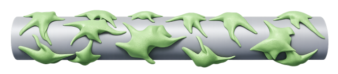
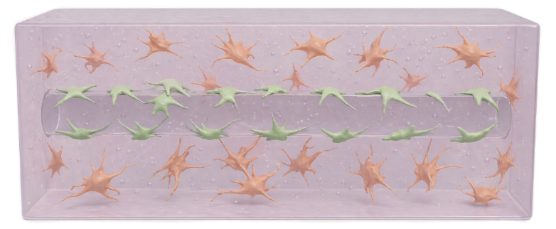
Cells can grow directly on the template before embedding, or be seeded later. A simple cool-down to 4 °C dissolves the polymer within minutes, leaving cell-lined, perfusable channels behind.
No toxic solvents. No cell damage. No expensive equipment. Plug and play.
Three steps, no specialized hardware, no post-seeding or pre-treatment.
An OXA template is added to the chip. Cells can optionally be grown and attached to its surface beforehand, or seeded later.

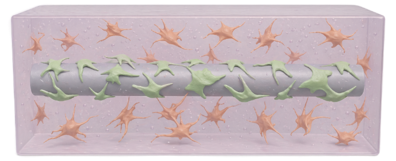
Hydrogel containing your cells is cast around the template and crosslinked, locking the geometry in place.
A short cool-down in the fridge dissolves the template in minutes, leaving a clean, cell-lined, perfusable channel.
Designed to drop into existing workflows and reach the resolution real tissue requires.
Compatible across a broad range of matrices.
Plug & play. No bioprinter or specialized hardware.
No post-seeding or pre-treatment required.
Fine enough to replicate capillaries and axonal guides.
Demonstrated across multiple physiologically relevant tissue models.
Endothelial cells and astrocytes arranged in physiologically correct geometry.
Endothelial-lined channels with supporting stromal cells.
Schwann-cell conduits guiding human neuron axon growth across channels.
From drug screening to disease modeling to next-generation chip design.
Drug screening and preclinical testing with more predictive human models, reducing reliance on animal studies.
Disease modeling, tissue microenvironment studies, and tissue innervation in a reproducible format.
Integrate with and improve current chips, enabling rich hydrogel architectures.
We're currently accepting pilot collaborations with pharma, biotech, academic, and organ-on-chip teams.
Get in touch